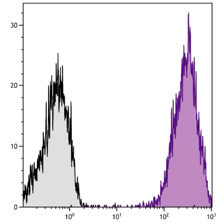
Human CD44 Antibody : APC

Katalog-Nummer OASB00953
Size : 100tests
Marke : Aviva Systems Biology
Contact local distributor :
 Telefonnummer : +1 850 650 7790
Human CD44 Antibody : APC (OASB00953)
| Datasheets/Manuals | Printable datasheet for Human CD44 Antibody : APC (OASB00953) |
|---|
| Tested Species Reactivity | Human |
|---|---|
| Predicted Species Reactivity | Human |
| Clonality | Monoclonal |
| Clone | F10-44-2 |
| Isotype | IgG2a |
| Host | Mouse |
| Conjugation | APC |
| Application | FC |
| Additional Information | Description: CD44, also known as phagocytic glycoprotein-1 (Pgp-1), exists as a large number of different isoforms resulting from alternative RNA splicing. The major isoform expressed on lymphocytes, myeloid cells, and erythrocytes is a glycosylated type I transmembrane protein. Other variable isoforms containing glycosaminoglycans have molecular weights ranging from 110 to 250 kDa and are widely expressed on hematopoietic and non-hematopoietic cells. CD44 contributes to the adhesion of leukocytes to endothelial cells, stromal cells, and the extracellular matrix. It also plays a functional role in cell migration, lymphocyte homing, and adhesion during hematopoiesis and lymphocyte activation. |
| Reconstitution and Storage | Store at 2-8C |
| Immunogen | Purified human T cells |
| Concentration | Lot specific |
| Specificity | CD44 |
| Characterization | To insure lot- to- lot consistency, each batch of product is tested by flow cytometry to conform tocharacteristics of a standard reference reagent. |
| Warning | Reagents contain sodium azide which is very toxic if ingested or inhaled. Avoid contact with skin, eyes, orclothing. Wear eye or face protection when handling. If skin or eye contact occurs, wash with copiousamounts of water. If ingested or inhaled, contact a physician immediately. Sodium azide yields toxichydrazoic acid under acidic conditions. Dilute azide- containing compounds in running water beforediscarding to avoid accumulation of potentially explosive deposits in lead or copper plumbing. |
| Dilution | Flow Cytometry: Purified (UNLB) antibody Fluorescein conjugate Biotin conjugate PE, APC, PE/CY7, and SPRD conjugates <= 1 ug/106 cells 10 uL/106 cells 10 uL/106 cells 10 uL/106 cells |
| Application Info | Flow cytometry, Immunohistochemistry (frozen sections8 and paraffin sections7), Immunoprecipitation |
| Other Applications Data | Since applications vary, you should determine the optimum working dilutionof the product that is appropriate for your specific need. |
| Storage | - The purified (UNLB) antibody is supplied as 0.1 mg of purified immunoglobulin in 1.0 mL of 100 mMborate buffered saline, pH 8.2. No preservatives or amine- containing buffer salts added. Store at 2- 8 C - The fluorescein (FITC) conjugate is supplied as 100 tests in 1.0 mL of PBS/NaN3. Store at 2- 8 C - The biotin (BIOT) conjugate is supplied as 100 tests in 1.0 mL of PBS/NaN3. Store at 2- 8 C - The R- phycoerythrin (R- PE), allophycocyanin (APC), and Spectral Red (SPRD) conjugates aresupplied as 100 tests in 1.0 mL of PBS/NaN3 and a stabilizing agent. Store at 2- 8 C. Do not freeze! - The R- phycoerythrin- Cyanine 7 (R- PE- CY 7) conjugates are supplied as 100 tests in 1.0 mL ofPBS/NaN3 and a stabilizing agent. Store at 2- 8 C. Do not freeze! - Protect conjugated forms from light. Reagents are stable for the period shown on the label if stored asdirected. |
| Reference | 1. McMichael, A.K., P.C.L. Beverly, S. Cobbold, M.J. Crumpton, W. Gilks, F.M. Gotch, N. Hogg, M. Horton, N. Ling, I.C.M. MacLennan, D.Y Mason, C. Milstein, D. Spiegelhalter, and H. Waldmann, eds. 1987. Leukocyte Typing III: White Cell Differentiation Antigens, OxfordUniversity Press, Oxford 2. Knapp, W., B. Dorken, W.R. GIlks, E.P. Rieber, R.E. Schmidt, H. Stein, A.E.G.K. Von dem Borne, eds. 1989. Leukocyte Typing IV: WhiteCell Differentiation Antigens, Oxford University Press, Oxford 3. Schlossman, S., L. Bloumsell, W. Gilks, J.M. Harlan, C. Kishimoto, J. Ritz, S. Shaw, R. Silverstein, T. Springer, T.F. Tedder, and R.F. Todd,eds. 1995. Leukocyte Typing V: White Cell Differentiation Antigens, Oxford University Press, Oxford 4. Barclay, A.N., M.H. Brown, S.K.A. Law, A.J. McKnight, M.G. Tomlinson, and P.A. van der Merwe, eds. 1997. The Leukocyte AntigensFacts Book, 2nd Edition, CD44 Section, Academic Press, New York, p. 240 5. Goldstein, L.A., and E.C. Butcher. 1990. Immunogenetics 32:389 6. Lesley, J., R. Hyman, and P.W. Kincade 1993. Adv. Immunol. 54:271 7. Anwar, F., and B. Wood. 2000. Mod Pathol. 13(10):1121- 1127 8. Ellis, P.A., Hart, D.N.J, Colls, B.M, Nimmo, J.C., MacDonald, J.E., and H.B. Angus. 1992. Clin Exp Immunol 90, 117- 123. |
|---|---|
| Gene Symbol | CD44 |
| Gene Full Name | CD44 antigen |
| Alias Symbols | IN, LHR, MC56, MDU2, MDU3, MIC4, Pgp1, CDW44, CSPG8, HCELL, HUTCH-I, ECMR-III |
| NCBI Gene Id | 960; 105717277 |
| Protein Name | CD44 antigen |
| Description of Target | The protein encoded by this gene is a cell-surface glycoprotein involved in cell-cell interactions, cell adhesion and migration. It is a receptor for hyaluronic acid (HA) and can also interact with other ligands, such as osteopontin, collagens, and matrix metalloproteinases (MMPs). This protein participates in a wide variety of cellular functions including lymphocyte activation, recirculation and homing, hematopoiesis, and tumor metastasis. Transcripts for this gene undergo complex alternative splicing that results in many functionally distinct isoforms, however, the full length nature of some of these variants has not been determined. Alternative splicing is the basis for the structural and functional diversity of this protein, and may be related to tumor metastasis. |
| Uniprot ID | P16070, A0A2K5DU87 |
| Protein Accession # | NP_000601.3 |
| Nucleotide Accession # | NM_000610.3 |