F Antibody : FITC

Katalog-Nummer OABF00317-FITC
Size : 100ul
Marke : Aviva Systems Biology
Contact local distributor :
 Telefonnummer : +1 850 650 7790
F Antibody : FITC (OABF00317-FITC)
Click here to learn more about Aviva's By-Request Conjugation Service.
| Datasheets/Manuals | Printable datasheet for OABF00317-FITC |
|---|
| Predicted Species Reactivity | Measles virus |
|---|---|
| Product Format | Liquid. Purified antibody supplied in 1x PBS buffer. |
| Clonality | Polyclonal |
| Isotype | IgG |
| Host | Rabbit |
| Conjugation | FITC: Fluorescein Isothiocyanate |
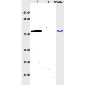
| Application | WB, IHC-P, IF |
| Additional Information | Subcellular location: Cytoplasm |
| Reconstitution and Storage | All conjugated antibodies should be stored in light-protected vials or covered with a light protecting material (i.e. aluminum foil). Conjugated antibodies are stable for at least 12 months at 4C. If longer storage is desired (24 months), conjugates may be diluted with up to 50% glycerol and stored at -20C to -80C. Freezing and thawing conjugated antibodies will compromise enzyme activity as well as antibody binding. |
| Immunogen | KLH conjugated synthetic peptide derived from Measles virus fusion protein |
| Purification | Purified by Protein A. |
| Concentration | 1 mg/ml |
| Application Info | WB(1:100-1000) IHC-P(1:100-500) IF(1:50-200) |
| Gene Symbol | F |
|---|---|
| Alias Symbols | Fusion glycoprotein F, F |
| NCBI Gene Id | 1489800 |
| Description of Target | Class I viral fusion protein. Under the current model, the protein has at least 3 conformational states: pre-fusion native state, pre-hairpin intermediate state, and post-fusion hairpin state. During viral and plasma cell membrane fusion, the heptad repeat (HR) regions assume a trimer-of-hairpins structure, positioning the fusion peptide in close proximity to the C-terminal region of the ectodomain. The formation of this structure appears to drive apposition and subsequent fusion of viral and plasma cell membranes. Directs fusion of viral and cellular membranes leading to delivery of the nucleocapsid into the cytoplasm. This fusion is pH independent and occurs directly at the outer cell membrane. The trimer of F1-F2 (F protein) probably interacts with H at the virion surface. Upon HN binding to its cellular receptor, the hydrophobic fusion peptide is unmasked and interacts with the cellular membrane, inducing the fusion between cell and virion membranes. Later in infection, F proteins expressed at the plasma membrane of infected cells could mediate fusion with adjacent cells to form syncytia, a cytopathic effect that could lead to tissue necrosis (By similarity). |
| Uniprot ID | P69353 |